Echocardiography (Trans-Thoracic and Trans-Esophageal Echocardiography)
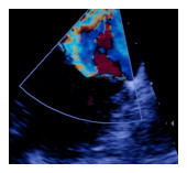 An echocardiogram is a diagnostic procedure that uses ultrasound waves
to produce a sonogram of the heart. Physicians use the images to measure
cardiac output, as well as the size and shape of the heart and how well
blood is flowing through the heart’s valves and chambers. A standard
echocardiogram is known as a trans-thoracic echocardiogram, or TTE. This
test is non-invasive and fast, and imaging is acquired externally
through a probe placed on the chest wall. An echocardiogram is a diagnostic procedure that uses ultrasound waves
to produce a sonogram of the heart. Physicians use the images to measure
cardiac output, as well as the size and shape of the heart and how well
blood is flowing through the heart’s valves and chambers. A standard
echocardiogram is known as a trans-thoracic echocardiogram, or TTE. This
test is non-invasive and fast, and imaging is acquired externally
through a probe placed on the chest wall.
In some cases, a physician
will order an alternative type of echocardiogram known as a
trans-esophageal echocardiogram, or TEE. An ultrasound probe is placed
into the patient’s esophagus – often after administration of a numbing
agent or while under conscious sedation. The probe is positioned behind
the heart, where it produces ultrasound views of the chambers and valves
not seen on traditional echocardiograms.
Back to Patient Education
|